Diese Arbeit stellt eine vergleichende Untersuchung von Pilzen, Mycetozoen (Schleimschimmelpilzen) und Bakterien vor, wobei deren biologische und morphologische Eigenschaften untersucht werden. Sie untersucht deren Entwicklung, Struktur und Klassifizierung, wobei der Schwerpunkt auf dem Lebenszyklus und den zellulären Merkmalen jeder Gruppe liegt. Die Studie trägt zu einem tieferen Verständnis der Pflanzenmorphologie und mikrobiellen Biologie bei, insbesondere in Bezug auf Pilze und Schleimschimmelpilze.
1810 x 1381 px | 30,6 x 23,4 cm | 12,1 x 9,2 inches | 150dpi
Weitere Informationen:
Dieses Bild ist ein gemeinfreies Bild. Dies bedeutet, dass entweder das Urheberrecht dafür abgelaufen ist oder der Inhaber des Bildes auf sein Urheberrecht verzichtet hat. Alamy berechnet Ihnen eine Gebühr für den Zugriff auf die hochauflösende Kopie des Bildes.
Dieses Bild kann kleinere Mängel aufweisen, da es sich um ein historisches Bild oder ein Reportagebild handel
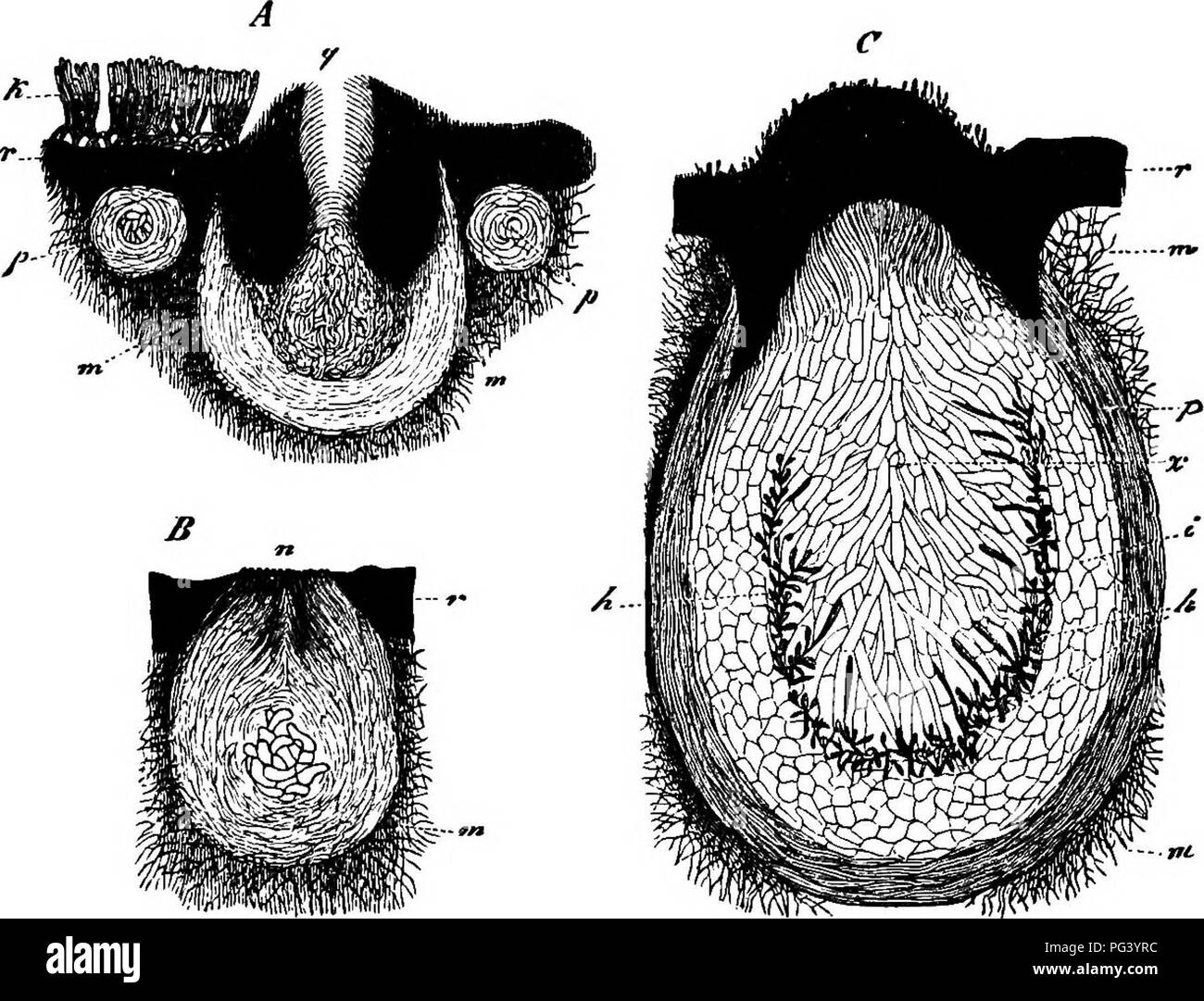
. Comparative morphology and biology of the fungi, mycetozoa and bacteria . Plant morphology; Fungi; Myxomycetes; Bacteriology. 2l6 DIVISION II.—COURSE OF DEVELOPMENT OF FUNGI. no longer be recognised, so that the perithecium in the mature state is broadly ovoido-conical with an indistinct ostiole. The wall is formed of three or four layers of not much thickened elongated cells. Phyllachora TJlmi appears to show similarity to the process here described. II. The club-shaped stroma of Xylariapolymorpha (Fig. 103) consists in the young state, according to my earlier observations, of a white medulla surrounded by a firm black rind. The former is composed of an air-containing tissue of colourless hyphae; the rind of the portion bearing perithecia consists of small-celled pseudo-parenchymatous tissue, which is overlaid on the outside by the hymenium which bears gonidia (see section LXXI) and ultimately disappears. The primordia. FIG. 103. Xylaria fiolyincrfiha. j4, B, C transverse sections through young stromata with perithecia divided more or less exactly in half, all three magn. go times, r rind, trt medullary layer of the stroma. A, p very young perithecium cut through the middle, p a similar one cut through near the median plane, (/ older perithecia, k gonidial layer. B perithecium with the mouth n bursting through the rind. C a nearly fuUy developed perithecium; the section passes close to the mouth, which is fashioned as at qi^A, elsewhere through the median plane; p the outer, i the inner wall of the perithecium, X the large.celled paraphyses filling the centre of the perithecium having entirely displaced the short-lived inner tissue, h the inner surface of the wall with the insertions of the paraphyses and asci. of the perithecia {A, p) make their appearance in the form of small spherical bodies which lie in the medulla close beneath the black rind, and are at once distinguished from the medullary tissue by containing no air and therefore being transparent. The