Ein Handbuch der Anatomie. Er Fasern continueon die gleiche Seite durch die dorsale Säule zu den Kernen cuneatusand gracilis. Von hier aus vermitteln die Fasern des zweiten Neurons die IN DER ATMUNG betroffenen TRAKTE 425 Impulse über den gegenüberliegenden medialen Lemniscus zum Thalamus.Das dritte Neuron verbindet den Thalamus mit dem kortikalen Bereich.Einige Fasern gehen von den Kernen gracilis und cuneatus zum cere-bellaren Cortex über; Neue Fasern gehen von hier in den dentaten Kern des Kleinhirns über, aus dem neue Fasern über die Bindehaut in den Thalamus gelangen. Ein Teil der Fasern, nur der ersten Neu
1291 x 1935 px | 21,9 x 32,8 cm | 8,6 x 12,9 inches | 150dpi
Weitere Informationen:
Dieses Bild ist ein gemeinfreies Bild. Dies bedeutet, dass entweder das Urheberrecht dafür abgelaufen ist oder der Inhaber des Bildes auf sein Urheberrecht verzichtet hat. Alamy berechnet Ihnen eine Gebühr für den Zugriff auf die hochauflösende Kopie des Bildes.
Dieses Bild kann kleinere Mängel aufweisen, da es sich um ein historisches Bild oder ein Reportagebild handel
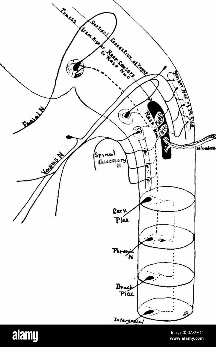
A manual of anatomy . he fibers continueon the same side through the dorsal column to the nuclei cuneatusand gracilis. From here the fibers of the second neuron convey the TRACTS CONCERNED IN RESPIRATION 425 impulses by way of the opposite medial lemniscus to the thalamus.The third neuron connects the thalamus with the cortical area.Some fibers pass from the nuclei gracilis and cuneatus to the cere-bellar cortex; new fibers pass from here to the dentate nucleus ofthe cerebellum from which new fibers pass to the thalamus throughthe brachia conjunctiva. Some of the fibers, only, of the first neuron, have the above course.Others, after entering the dorsal roots of the spinal nerves, do notenter the dorsal column but join the spinocerebellar tracts (ventraland dorsal superficial) to end in the cerebellar cortex of thesan-ie side. The impulses are then carried to the dentate nucleusand from here through the brachium conjunctivum to the thalamus;from the thalamus the impulses are conveyed to the cerebral cortex.. Flc. 309.— Diagram of the nerves and tracts concerned in respiration. Respiration.—Although respiration is apparently controlled bythe respirator)^ nucleus that lies in the formatio reticularis of theoblongata, it is maintained by stimuli carried to this center by theblood vascular system and reflex impulses from the sensor portionof the vagus through cells in the nucleus of termination of the vagalnerve and by impulses from the higher respiratory centers. Therespiratory nucleus is connected with the following motor nuclei:Facial, vagal, accessory, cervical plexus, phrenic, brachial plexusand thoracic nerves. Axones from the higher centers and from the 426 THE NERVE SYSTEM sensor vagal nucleus end in the respiratory nucleus. The cells of therespiratory nucleus send their axones directly, or by means of col-laterals, in the formatio reticularis, to the nuclei of the above-mentioned motor nerves so that through this connection a numberof cerebral and spinal ner