Eine elektronenmikroskopische Zytologie-Untersuchung zur Untersuchung der submikroskopischen Struktur des Zytoplasmas. Zu den wichtigsten Erkenntnissen gehört die Identifizierung des endoplasmatischen Retikulums und seine Unterscheidung in glatte und raue Typen.
1848 x 1352 px | 31,3 x 22,9 cm | 12,3 x 9 inches | 150dpi
Weitere Informationen:
Dieses Bild ist ein gemeinfreies Bild. Dies bedeutet, dass entweder das Urheberrecht dafür abgelaufen ist oder der Inhaber des Bildes auf sein Urheberrecht verzichtet hat. Alamy berechnet Ihnen eine Gebühr für den Zugriff auf die hochauflösende Kopie des Bildes.
Dieses Bild kann kleinere Mängel aufweisen, da es sich um ein historisches Bild oder ein Reportagebild handel
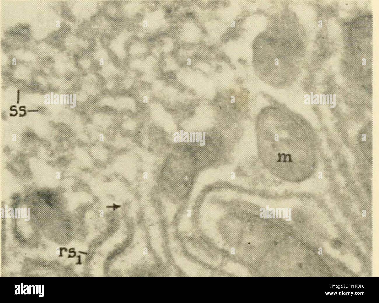
. Cytology. Cytology. The electron microscope studies published to date on the submicro- scopic structure of the cytoplasm are more or less in general agreement concerning the reality of the endoplasmic reticulum, but considerable. r' Figure 3-24. Electron Micrograph of Portion of Cytoplasm of Rat Parenchymal Liver Cell. Note local differentiation of membrane elements of the endoplasmic reticulum into smooth- and rough-surfaced types. The smooth-surfaced elements (ss) appear as vesicles and tubules disposed in discontinuous groups mostly at the periphery of the cell. The rough-surfaced elements (rsj rso) are arranged more or less parallel to one another in large arrays. Profiles of rough-surfaced elements sectioned normal to their surface are marked as rsj, and oblique to their surface as rso. Points of continuity between the smooth- and rough-surfaced elements are indicated by arrows. Numerous mitochondria (m) are seen in the field. (From Palade, G. E., 1956. "The Endoplasmic Reticulum, " J. Biophys. Biochem. CytoL, 2, Suppl., Fig. 3, Plate 33.) confusion still exists regarding terminology and interpretation of indi- vidual components of the system. For example, the cisternae are inter- preted by Porter and Palade as pairs of membranes arranged in parallel and connected at their ends. Sjostrand (1956), on the other hand, suggests that the cisternae actually represent a discontinuous series of STRUCTURE AND FUNCTION OF CYTOPLASMIC ORGANELLES / 53. Please note that these images are extracted from scanned page images that may have been digitally enhanced for readability - coloration and appearance of these illustrations may not perfectly resemble the original work.. Wilson, G. B. (George Bernard), 1914-; Morrison, John H. (John Herbert), 1927-. New York, Reinhold