. Eine praktische Abhandlung über die medizinische Diagnose für Studenten und Ärzte . der Medianlinie. Die Pulsierung der Leber, die vaskulären Ursprungs und damit synchron mit der Herzpulsation ist, ist im Leberbereich bei der Erweiterung des rechten Herzens seenin. Magen- und Darmbewegungen. PeristaMc Bewegung, ob der Magen, oder des großen oder Dünndarms, kann durch die Bauchwände sichtbar sein. Bei der Magendilatation und Gastroptose kann man die Wellen in rhythmischer Abfolge von links nach rechts im Bauchraum beobachten. Ihr allgemeiner Kurs ist normalerweise von den Leftup
2050 x 1219 px | 34,7 x 20,6 cm | 13,7 x 8,1 inches | 150dpi
Weitere Informationen:
Dieses Bild kann kleinere Mängel aufweisen, da es sich um ein historisches Bild oder ein Reportagebild handel
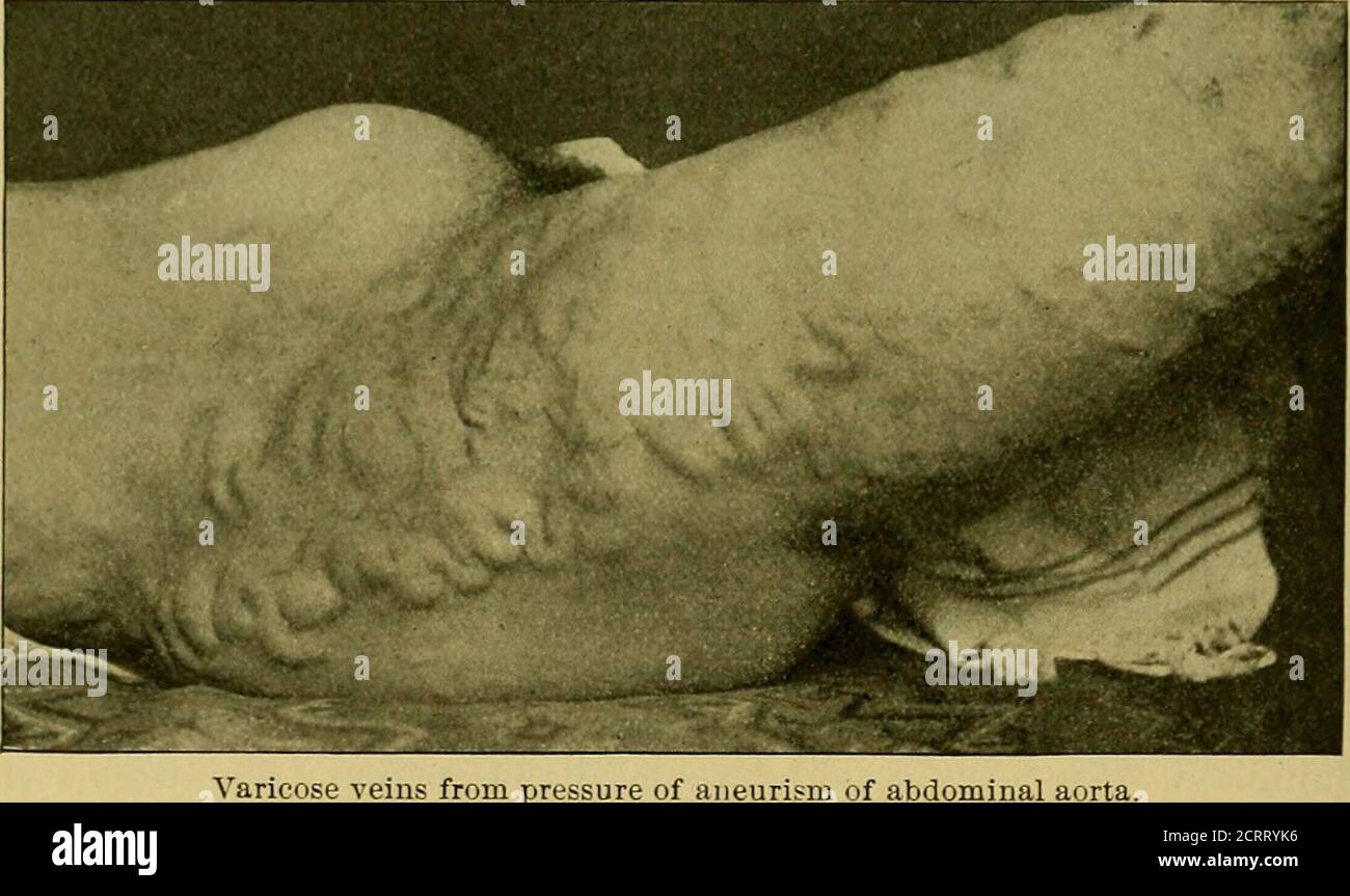
. A practical treatise on medical diagnosis for students and physicians . of the median line. Pulsation of the liver, which is ofvascular origin and therefore synchronous with cardiac pulsation, is seenin the hepatic area in dilatation of the right heart. Gastric and Intestinal Movements. PeristaMc movement, whether ofthe stomach, or of the large or small intestine, may be visible throughthe abdominal walls. In gastric dilatation and gastroptosis the wavesmay be seen in rhythmical succession passing from Left to right in thecentre of the abdomen. Their general course is usually from the Leftupper to the right lower quadrant. When due to movements of thelarge intestine, the waves follow the course of the canal ; while thosewhich emanate from the small intestine are confined to the region aroundthe umbilicus. Visible peristalsis when of gastric origin indicatesobstruction at the pylorus; intestinal peristalsis is seen when the lumenof the bowel is obstructed. The movements may be excited by fillipingthe abdomen with a towel wrung out of cold water. Fig. 183.. of abdominal aorta. pe. In general enlargement of the abdomen the shape is uniform.In very fat subjects and in women with relaxed abdominal walls theabdomen may be pendulous. (See Fig. 182). In ascites the tissue overthe umbilicus may protrude and form a localized prominence in womenwhose abdominal walls have previously been relaxed. Abdominal en-largements due to ascites sometimes assume a peculiar cone-shape (seeAbdominal Walls), the base corresponding to the plane of the abdo-men, the apex protruding below the umbilicus ; this is particularly thecase when the patient has had to maintain the semi-erect position for sometime. Local enlargements, such as morbid growths or changes in the sizeof viscera, produce irregularities in the surface corresponding in positionto the internal lesion. The shape varies momentarily in hysterical dis- PALPATION AND PERCUSSIONOF THE ABDOMEN 509 tention. In wasting disease of the v