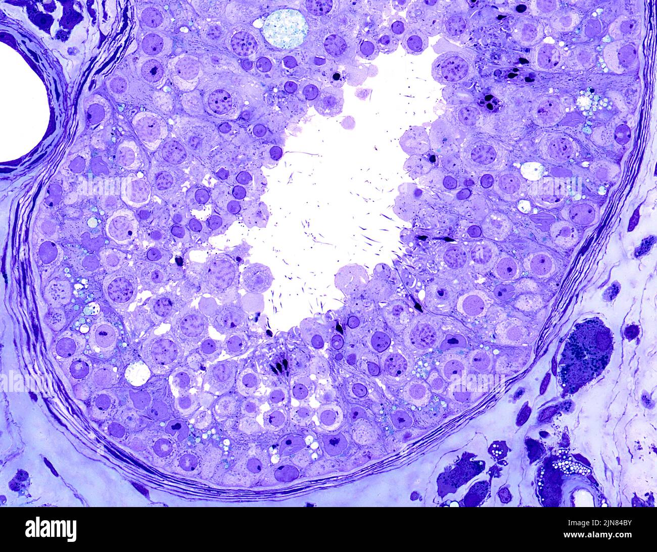
Human testis, light micrograph. Seminiferous tubule of the human testis showing the seminiferous epithelium. All types of germ cells are present representing the process of spermatogenesis. Mature spermatids with a head and tail are seen. From stem cell spermatogonia to fully-formed spermatids requires 70 days in humans. Cells with irregular shaped nuclei are the non-dividing Sertoli cells. The peritubular tissue boundary of the tubule contains fibroblasts and contractile smooth muscle myoid cells. Epoxy resin section, Toluidine blue stain. Magnification: x310 when width printed at 10cm.