. Pathogene Mikroorganismen. Ein text-Buch der Mikrobiologie für Ärzte und Studenten der Medizin. (Auf Williams' Bakteriologie). Bakteriologie; pathogenen Bakterien. 424 SPEZIFISCHE MIKROORGANISMEN andere Einzelpersonen. Die Zelle ist 8 bis 60/i im Durchmesser. Die ecto - keimplasma ist deutlich von der endoplasm differenziert, auch wenn die Zelle unbeweglich ist, und der lobose pseudopodia sind völlig von Stiff hoch refraktive Ektoplasma. Die endopla' fem enthält Essen Material bestehend aus Bakterien, Zelle, Fragmente und roten Blutkörperchen. Der Kern ist sehr deutlich sichtbar in der lebenden Amöben
1743 x 1434 px | 29,5 x 24,3 cm | 11,6 x 9,6 inches | 150dpi
Weitere Informationen:
Dieses Bild ist ein gemeinfreies Bild. Dies bedeutet, dass entweder das Urheberrecht dafür abgelaufen ist oder der Inhaber des Bildes auf sein Urheberrecht verzichtet hat. Alamy berechnet Ihnen eine Gebühr für den Zugriff auf die hochauflösende Kopie des Bildes.
Dieses Bild kann kleinere Mängel aufweisen, da es sich um ein historisches Bild oder ein Reportagebild handel
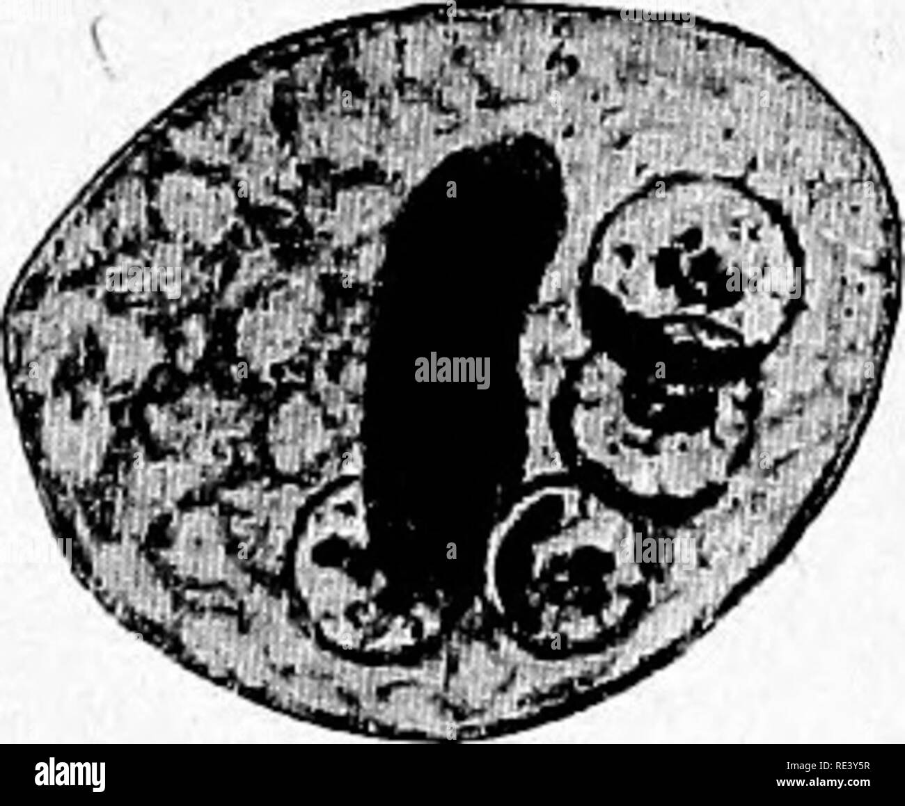
. Pathogenic micro-organisms. A text-book of microbiology for physicians and students of medicine. (Based upon Williams' Bacteriology). Bacteriology; Pathogenic bacteria. 424 SPECIFIC MICRO-ORGANISMS Other individuals. The cell is 8 to 60/i in diameter. The ecto- plasm is distinctly differentiated from the endoplasm even when the cell is motionless, and the lobose pseudopodia are made up entirely of the stiff highly refractive ectoplasm. _The endopla'fem contains food material consisting of bacteria, cell fragments and red blood cells. The nucleus is very distinctly visible in the living ameba. It is spherical and surrounded by a-thick doubly, contoured nuclear membrane. The chromatin is usually dis- tributed just beneath the nuclear membrane in largest amount and in the center there is a karyosome with definite centriole. The vege- tative multiplication takes place by division into two daughter cells. Multiple division seems not to occur. Pig. 184.—Enda- ^ , . , , i rri i. mceba dysenteria. Lyst formation IS rarely observcd. 1 he cysts 5ntgfoTfnuTeI ^re most likely to be found when the stool be- and a mass of chro- comes formed ill convalcsccncc from an attack of midial substance. i . , i . i ^i -u {After Hartmann.) dysentery and they may then be very numer- * ous. The mature cyst contains four nuclei, , and frequently contains also one or more large masses of chromidial substance which stain black with iron hematoxylin. The forms of the organism commonly observed in the feces of dysentery are either the active vegetative cells^ or degenerating forms, and the latter may lead to confusion unless their true nature is recognized. The belief that amebae bear a causal relation to dysentery is based upon the fact that certain types of amebae, E. dysmtericB (E. Mstolyiica) are found in the stools, as a rule, only in cases of dysentery; further, that these cases of dysentery, in which these amebae occur, are characterized by definite clinical signs and typical anatomical