5549 x 2551 px | 47 x 21,6 cm | 18,5 x 8,5 inches | 300dpi
Aufnahmedatum:
1. Oktober 2014
Weitere Informationen:
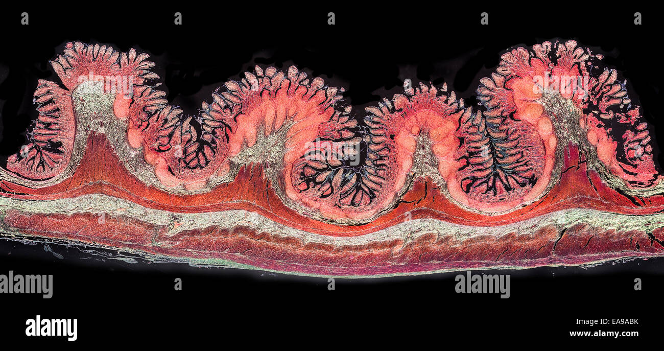
Intestinal villi (singular: villus) are small, finger-like projections that protrude from the epithelial lining of the intestinal wall. Each villus is approximately 0.5-1.6 mm in length, and has many microvilli projecting from the enterocytes of its epithelium which collectively form the striated or Brush border. Each of these microvilli are much smaller than a single villus. The intestinal villi should not be confused with the larger folds of mucous membrane in the bowel known as the plicae circulares. A villus is much smaller than a single fold of plicae circulares. Villi increase the internal surface area of the intestinal walls. Increased surface area allows for increased intestinal wall area that is available for absorption. Increased absorptive area is useful because digested nutrients (including monosaccharide and amino acids) pass into the semipermeable villi through diffusion, which is effective only at short distances. In other words, increased surface area (in contact with the fluid in the lumen) decreases the average distance travelled by nutrient molecules, so effectiveness of diffusion increases. The villi are connected to the blood vessels so the circulating blood then carries these nutrients away